Review Article 
 Creative Commons, CC-BY
The Use of Scientific Instruments in The Exploration of The View Plane in Drawing
*Corresponding author:John Robert Burns, John Burns Illustration, 54 Holmfield Road Coventry, West Midlands, CV2 4DE, UK.
Received:November 23, 2022; Published:January 24, 2023
DOI: 10.34297/AJBSR.2023.17.002414
Abstract
Keywords: Scientific instrument; Viewpoint; Picture Plane; Cone of Vision
Review
During the years of practice as a scientific illustrator - often dealing with setting up scenes that require a microscopic, extremely local point of view – I have had been required to employ specialist viewing facilities and instruments in order to study the close-up, microscopic field in which important but extremely small-scale events take place. Such events may be at the molecular, microscopic and micro-organism level. This has often required the use of specialist viewing instruments; sometimes using very narrow fields of visible light: at other times functioning in the x-ray, ultra-sound and electron based realm. Whilst such use of specialist, close-up viewing is very often seen as performing a supportive function in allowing for the visual scrutiny of microscopic events and the tiny but critical protagonists involved in those events at that scale, and the aesthetic qualities of the finished illustrative artwork are assessed within a more usual, human eyesight configuration - that is with cones of vision, focus and field coverage more readily associated with un-aided human observation – it is noticeable that the microscopic and close-up aspects of specialist instrument viewing have aesthetic qualities of their own.
There has lately been evidence of a growing interest in the perceived aesthetic qualities of close-up and time-based photography and filmed sequences of chemical and physical events happening at extremely small scale.
In the area of investigation that I have been engaging with here, there is some reference to the microscopic view of subject matter, but in this case the subject matter is the finished artwork itself: viewed not from the usual human eye point of view, but from the much closer, microscopic viewpoint of the scientific instrument that provided assistance in the creation of the artefact to be seen from the human eye viewpoint; in effect, creating a viewing loop in which the view of subject matter and resultant artwork are both seen using broad and extremely narrow cones of vision.
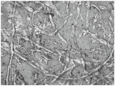
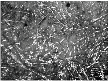
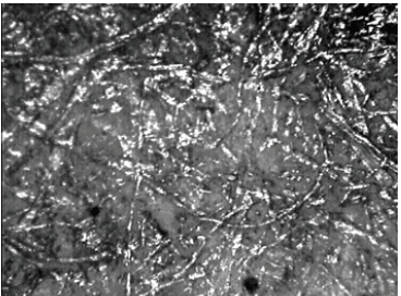
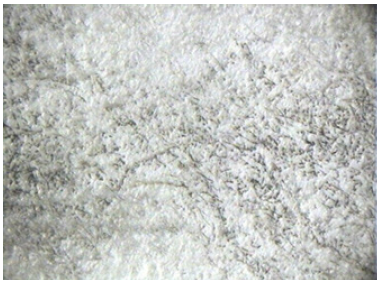
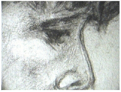
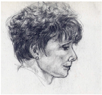
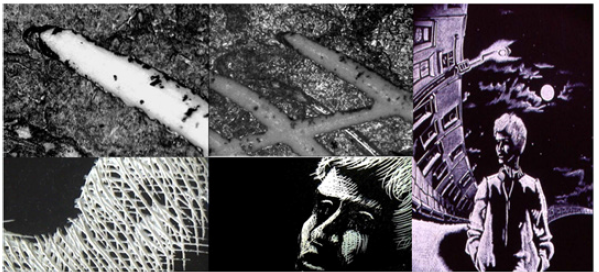
One of the interesting features of the microscopic to close-up viewing of drawn, painted or photographed imagery is the point at which graphite, pigment, photo-emulsion grain or digital picture elements cross the awareness boundary, ceasing to be abstract arrangements – as far as the viewer is concerned – and become recognisable as arrangements of marks and shapes that hint that the viewer may have seen something like this before. Of course, an arrangement of graphite smears on a fibrous background is not an image of a face; it is rather an arrangement of just that, graphite smears that absorb and reflect photons in different directions, thereby making the surface where they lie seem to be darker than the surrounding surface. When seen from a certain viewpoint, using a cone-of-vision as normally associated with the human eye, these reflecting and deflecting smears can appear to hint that, in the wider outside world, we may have seen hints and shades arranged in a similar way and which we recognise as being a visual indication of other persons, beings and scenes (Figure 1-7).



 We use cookies to ensure you get the best experience on our website.